Mogedda E Haiba1 ![]() ,
Ebtehal S Al-Abdullah2,
Hazem A Ghabbour2,3,
Sayed M Riyadh4,5,
Reham M Abdel-Kader5
,
Ebtehal S Al-Abdullah2,
Hazem A Ghabbour2,3,
Sayed M Riyadh4,5,
Reham M Abdel-Kader5
For correspondence:- Mogedda Haiba Email: mogedda.haiba@yahoo.com
Received: 19 April 2016 Accepted: 10 October 2016 Published: 28 November 2016
Citation: Haiba ME, Al-Abdullah ES, Ghabbour HA, Riyadh SM, Abdel-Kader RM. Inhibitory activity of benzo[h]quinoline and benzo[h]chromene in human glioblastoma cells. Trop J Pharm Res 2016; 15(11):2337-2343 doi: 10.4314/tjpr.v15i11.6
© 2016 The authors.
This is an Open Access article that uses a funding model which does not charge readers or their institutions for access and distributed under the terms of the Creative Commons Attribution License (http://creativecommons.org/licenses/by/4.0) and the Budapest Open Access Initiative (http://www.budapestopenaccessinitiative.org/read), which permit unrestricted use, distribution, and reproduction in any medium, provided the original work is properly credited..
Purpose: To carry out a neat synthesis of 2-amino-5,6-dihydro-8-methoxy-4-phenylbenzo[h]quinoline-3-carbonitrile (compound 2) and 2-amino-5,6-dihydro-8-methoxy-4-phenyl-4H-benzo[h]chromene-3-carbonitrile (compound 3) and evaluate their cytotoxic activity in human glioblastoma cells.
Methods: Benzo[h]quinoline and benzo[h]chromene were synthesized by treating 6-methoxy-1-tetralone with benzylidenemalononitrile under microwave irradiation. The structures of compounds 2 and 3 were confirmed by elemental, spectral, and x-ray crystallographic analyses. The cytotoxic activity of compounds 2 and 3 was evaluated using WST-1 assay in U373 human glioblastoma cell line.
Results: The molecular structures of compounds 2 and 3 were demonstrated unambiguously from single crystal x-ray measurements and they crystallized in triclinic form, P-1, for both compounds. In-vitro cytotoxic activity data for compound 2 in human glioblastoma cell line (U373) indicate that no significant cytotoxicity was observed. On the other hand, compound 3 showed highly significant cytotoxic effects on U373 cells at concentrations starting from 0.1 μg/ mL.
Conclusion: Compound 3 produces a decrease in cell viability with approximately 80 % cell death while compound 2 did not indicate significant cytotoxic activity. This suggests that the chromene moiety of compound 3 may be responsible for its high cytotoxicity.
Introduction
Literature survey has demonstrated the diversity of pharmacological applications of fused chromenes such as, anticancer [1,2], antibacterial [3], anti-inflammatory [4], antioxidants [5,6] and antiprotozoal [7] activities. Also, fused quinoline derivatives produce wide pharmacological activities such as antibacterial [8,9], antifungal [10], antimalarial [11], antiplasmodium [12] and anticancer [13,14] effects. Moreover, they have been reported to act as vesicular glutamate transporters [15] and HIV-Integrase inhibitors [16]. The combination of different ring systems in one compound is one of the major tools implemented in this study to produce more biologically active products in the field of drug development, aiming to obtain more active and less toxic products. Based on the above knowledge and in continuation of our previous work [17-19], the present study provides an evidence for the high efficient biological activity of the synthesized benzo[h]quinoline and benzo[h]chromene derivatives as anticancer compounds.
Methods
Materials and instruments
Melting points were recorded on a Barnstead 9001 Electrothermal melting point apparatus and are uncorrected. IR spectra were recorded on a Perkin Elmer FT-IR Spectrum BX Spectrometer at cm-1 scale using KBr discs. 1H-NMR and 13C-NMR were recorded on a JEOL 500 MHz Spectrometer, (Japan) and chemical shift values were expressed in δ values (ppm) relative to tetramethylsilane (TMS) as an internal standard. Coupling constants are given in Hz. The mass spectra were recorded on GCMC-QP 1000 EX Shimadzo Gas Chromatography MS spectrometer, (Japan) E.I. 70 ev. Elemental analysis (C, H, N) were carried out at the Micro analytical Center, Faculty of Science, Cairo University, Egypt, and were in full agreement with the proposed structures within ± 0.1-0.2 % of the theoretical values. All reagents were of analytical grade. The reactions was followed up by analytical thin layer chromatography (TLC) on pre-coated (0.75 mm) silica gel GF254 plates (E. Merck, Germany) and the products were visualized by UV light at 254 nm.
Synthesis of 2-amino-5,6-dihydro-8-methoxy-4-phenyl-benzo[h]quinoline-3-carbonitrile (2)
In a 5 mL glass tube, to a mixture of 6-methoxy-1-tetralone (1) (0.176 g, 0.001 mol), 2-benzylidenemalononitrile (0.154 g, 0.001 mol) in ethanol (3 mL), ammonium acetate (0.154 g, 0.002 mol), few drops of piperidine were added. The reaction tube was placed inside the cavity of the microwave synthesis system the tube was irradiated in the microwave oven with a power of 400 W, at a temperature of 130 oC, for 15 min. After completion of the reaction (monitored by TLC) and cooling, the solid formed was filtered off, washed with dilute cold ethyl alcohol and recrystallized from ethyl alcohol to give compound 2.
Synthesis of 2-amino-5,6-dihydro-8-methoxy-4-phenyl-4H-benzo[h]chromene-3-carbonitrile (3)
In a 5 mL glass tube, a mixture of 6-methoxy-1-tetralone (1) (0.176 g, 0.001 mol), 2-benzylidenemalononitrile (0.154 g, 0.001 mol) in ethanol (3 mL), few drops of piperidine were added. The reaction tube was placed inside the cavity of the microwave synthesis system, the tube was irradiated in the microwave oven with a power of 400 W, at a temperature of 130 oC, for 15 min. After completion of the reaction (monitored by TLC) and cooling, the solid formed was filtered off, washed with dilute cold ethyl alcohol and recrystallized from ethyl alcohol to yield compound 3.
X-ray crystallographic analysis
Compounds 2 and 3 were obtained as single crystals by slow evaporation from ethanol solution of the pure compound at room temperature. Data were collected on a Bruker APEX-II D8 Venture diffractometer, worked with graphite monochromatic Mo Kα radiation at 100 (2) K. Cell refinement and data reduction were carried out by Bruker SAINT. SHELXS-97 [20, 21] was used to solve the structures. The final refinement was carried out by full-matrix least-squares techniques with anisotropic thermal data for nonhydrogen atoms on 𝐹. CCDC 140449 and 1404416 contains the supplementary crystallographic data for these compounds and can be obtained free of charge from the Cambridge Crystallographic Data Centre via www.ccdc.cam.ac.uk/data_request/cif.
Cytotoxicity evaluation
U373 human glioblastoma cell line was used to evaluate the cytotoxicity of the compounds. U373 cells were maintained in DMEM medium containing 10 % fetal bovine serum and 1 % penicillin/streptomycin. Cells were seeded in 96 well plates at a density of 5000 cells/well in a final volume of 100 μL/well overnight at 37 °C and 5 % CO2. Compounds 2 and 3 were dissolved in DMSO and 5 different concentrations (0.1, 1, 10, 100 and 200 μg/mL) were prepared from each compound.
Drugs were added to the cells and the plates were incubated for an additional 48 hours at 37 °C and 5 % CO2. Control cells were incubated with DMSO only. Cell viability was assayed using WST-1 assay (Clontech) according to the manufacturer's protocol. Briefly, 10 µL of Premixed WST-1 Cell Proliferation Reagent was added to each well after drug incubation for 48 h and absorbance was measured 4 h later at 450 nm using Perkin Elmer Victor 1530 plate reader. Each experiment was performed in duplicates, and experiments were repeated for each concentration 3 or 4 times.
Statistical analysis
Data were analyzed using Prism 5.0 software (GraphPad Software, LaJolla, CA, USA). Statistical significance was assessed by one-way ANOVA test. Data are expressed as mean ± SEM. P < 0.05 was considered statistically significant.
Results
Subjecting a mixture of 6-methoxy-1-tetralone (1), benzylidenemalononitrile, ammonium acetate (in molar ratio 1:1:2, respectively) in ethanol, and drops of piperidine as a catalyst, to microwave irradiation afforded 2-amino-5,6-dihydro-8-methoxy-4-phenyl-benzo[h] quinoline-3-carbonitrile (2) as the isolated product (Scheme 1).
On the other hand, subjecting a similar amount of the mixture 6-methoxy-1-tetralone (1), benzylidenemalononitrile in ethanol and drops of piperidine to microwave irradiation under the same reaction conditions, however in the absence of ammonium acetate, furnished the respective 2-amino-5,6-dihydro-8-methoxy-4-phenyl-4H-benzo[h] chromene-3-carbonitrile (3) (cf. Scheme 1).
The structures of compounds 2 and 3 were confirmed by their elemental, spectral, (IR, 1H NMR, 13C NMR and MS) and x-ray crystallographic analyses.
2-Amino-5,6-dihydro-8-methoxy-4-phenyl-benzo[h]quinoline-3-carbonitrile (2)
Yield 80 %; m.p.: 222-224 oC; IR (υmax / cm-1): 3475, 3379 (NH2), 2208 (CN), 1625 (C=N); 1H-NMR (CDCl3): δ, 2.6 (m, 2H, CH2), 2.7 (m, 2H, CH2), 3.8 (s, 3H, OCH3 ), 5.1 (s, 2H, NH2), 6.7 (s, 1H, Ar, CH-7), 6.8 (d, J = 8.4 Hz, 1H, Ar-CH-9), 7.3 (m, 2H, Ar-CH-2`,6`), 7.4-7.5 (m, 3H, Ar-CH-3`,4`,5`), 8.2 (d, J = 8.4 Hz, 1H, Ar, CH-10); 13C-NMR (CDCl3): δ, 24.4 (CH2, C-5), 28.1 (CH2, C-6), 55.6 (OCH3), 88.09 (C-3), 113.05, 113.22, 117.53, 117.56, 126.6, 127.9, 128.8, 128.9, 129.1, 136.6, 141.6, 153.09, 154.6, 159.2, 161.3 (Ar-C and CN); MS: m/z (%): 327 (M+, 23.9) consistent with the molecular formula (C21H17N3O). Anal. Calcd. For C21H17N3O (327.13): C, 77.04; H, 5.23; N, 12.84. Found: C, 77.11; H, 5.44; N, 12.78 %.
2-Amino-5,6-dihydro-8-methoxy-4-phenyl-4H-benzo[h]chromene-3-carbonitrile (3)
Yield 85 %; m.p.: 226-228 oC; IR (υmax / cm-1): 3471, 3392 ( NH2), 2196 (CN), 1695 (C=N); 1H-NMR (DMSO-d6): δ: 1.79 (m, 1H, CH-5), 2.09 (m, 1H, CH-5), 2.55 (m, 1H, CH-6), 2.69 (m, 1H, CH-6), 3.72 (s, 3H, OCH3), 4.00 (s, 1H, CH-4), 6.73 (s, 1H, Ar-CH-7), 6.77-6.8 (m, 3H, Ar), 7.2-7.3 (m, 5H, Ar-H and NH2), 7.39 (d, J = 7.8 Hz, 1H, Ar-CH-10); 13C-NMR (DMSO-d6): δ, 24.7 (CH2-5), 27.5 (CH2-6), 42.7 (CH-4), 55.5 (OCH3), 56.8 (C-3), 109.2, 111.7, 113.9, 121.0, 121.5, 122.4, 127.4, 128.1, 129.0, 137.6, 140.2, 144.4, 159.6, 160.3 (Ar-C and CN); MS: m/z (%): 330 (M+, 29) consistent with the molecular formula (C21H18N2O2). Anal. Calcd. For C21H18N2O2 (330.13): C, 76.34; H, 5.49; N, 8.48. Found: C, 76.19; H, 5.61; N, 8.32 %.
X-ray
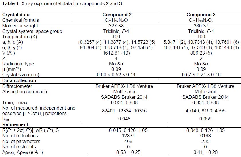
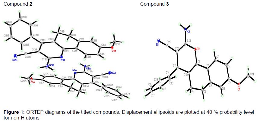
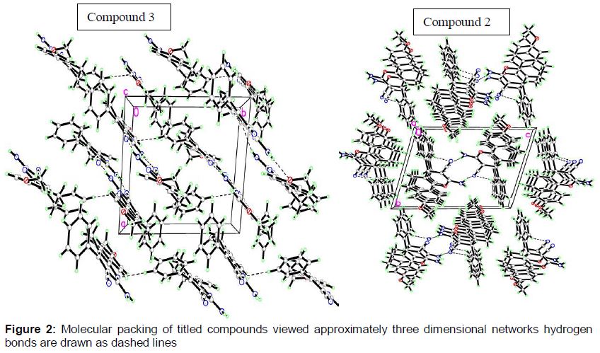
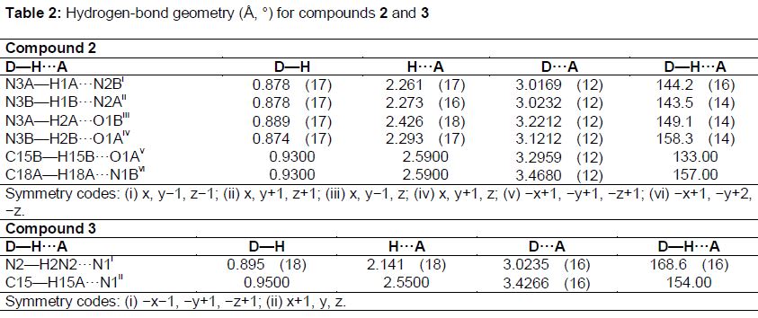
For the title compounds, C21H17N3O and C21H18N2O2, the crystallographic data and refinement information are summarized in . The asymmetric unit of compound 2 contains two independent molecules but in case of compound 3 it contains only one molecule as shown in . All the bond lengths and angles are in normal ranges [22]. In the crystal packing, , molecules of compound 2 are linked via six intermolecular hydrogen bonds (). Molecules of compound 3 are linked via two intermolecular hydrogen bonds ().
Cytotoxicity
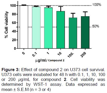
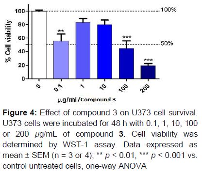
U373 human glioblastoma cell line was used for in-vitro cytotoxic screening of compounds 2 and 3, to evaluate their cytotoxicity at 5 different concentrations. Results declared that, compound 2 showed no significant cytotoxic effect on the U373 cells (). On the contrary, compound 3 showed significant cytotoxic effects on U373 cells starting at very low concentrations (0.1 μg/mL). Moreover, the highest concentration used in this study (200 μg/mL) led to a highly significant effect, decreasing cell viability to 20 % and causing approximately 80 % cell death ().
Discussion
The structures of benzo[h] quinoline-3-carbonitrile 2 and benzo[h]chromene-3-carbonitrile 3 have been established by x-ray crystallography and supported by spectral data. Mass spectra revealed a [M]+ ion peaks at 327 and 330 assignable to the molecular weight of compounds 2 and 3, respectively. In 1H NMR spectra characteristic signals resonating at δ = 5.1 ppm (for compound 2) and δ = 7.2-7.3 ppm (for compound 3) were typical for NH2 groups. IR showed absorption bands at 3475-3471, 3379-3392 (NH2), 2208, 2196 (CN) cm-1.
The anticancer activity results declared that compound 2 showed no significant cytotoxic effect on the U373 cells. Higher concentrations starting at 100 μg/mL demonstrated a slight non-significant decrease in cell viability compared to the controls (untreated cells). Incubating the U373 cells with 100 μg/mL of compound 2 for 48 h decreased cell viability to approximately 72 % compared to the control (), however, this effect was not statistically significant. On the other hand, compound 3 demonstrated high cytotoxic effects towards U373 human glioblastoma cell line. Incubation of U373 cells with 0.1 μg/mL of compound 3 decreased cell viability significantly to approximately 55 %, surprisingly increasing concentrations ranging from 1-10 μg/mL had a mild non-significant effect on cell viability (approximately 80 %). A remarkable cytotoxic effect was seen again at 100 μg/mL decreasing viability significantly to 45 %, and 200 μg/mL of compound 3 decreased cell viability by up to 20 %, meaning it led to approximately 80 % cell death ().
Conclusion
The present study provides a simple and rapid method for synthesizing benzo[h]quinoline 2 and benzo[h]chromene 3. The chromene moiety of compound 3 may be responsible for the high cytotoxicity, thus underlining the importance of the development of chromene derivatives in future work, with the aim to synthesize more active and less toxic products.
Declarations
Acknowledgement
References
Archives


News Updates